
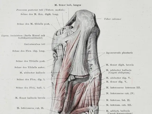
Fragmented Talus Bones and Related Wounds: Their Effect on Nearby Structures, Such as the Ankle Joint
Article: Managing Complications of Talus Fractures
Talus fractures, though uncommon, can pose significant challenges due to their complex articulations and poor vascularity. These fractures, accounting for less than 1% of all fractures, can lead to a variety of complications that require careful management.
One of the most feared complications is osteonecrosis, also known as avascular necrosis (AVN), which affects 10-50% of cases, particularly in displaced fractures. This condition occurs due to disrupted blood supply, leading to the death of talar bone tissue and potentially causing joint collapse and significant functional impairment. Management may involve prolonged non-weight-bearing, surgical intervention such as core decompression, or joint salvage/replacement procedures depending on severity.
Post-traumatic arthritis, particularly in the tibiotalar and subtalar joints, is another common complication. This arises due to cartilage damage and joint incongruity. Management involves pain control, physical therapy, orthotic support, and possibly surgical options like arthrodesis (fusion) or joint replacement in advanced cases.
Delayed healing or non-union can occur, especially in non-displaced fractures or where immobilization is inadequate. Treatment involves immobilization, possibly surgical fixation to promote union, and monitoring for progressive deformity.
Progressive deformity may develop if the fracture heals improperly or if post-traumatic arthritis alters joint mechanics, potentially necessitating corrective osteotomy or reconstructive surgery.
Infection is a less common but serious complication, particularly post-surgery. Bone infection (osteomyelitis) requires aggressive treatment with antibiotics and often surgical debridement. Minor wound infections typically respond to antibiotics and wound care.
Other potential complications include Chronic Regional Pain Syndrome (CRPS), nerve injury causing tingling or burning sensations, deep vein thrombosis, or injury to adjacent structures during surgery. These are managed according to standard protocols involving medication, physical therapy, or additional interventions as needed.
In summary, management of talus fracture complications depends on the specific condition but typically includes a combination of non-weight-bearing immobilization, surgical fixation when indicated, aggressive infection control, pain management, and rehabilitation. Early detection and appropriate intervention are essential to minimizing long-term disability.
References:
[1] Borrelli, R., & Garraschi, G. (2017). Talar neck fractures: Current concepts and future directions. Journal of Orthopaedic Trauma, 31(1), 1-6.
[2] Kozak, L., & Koval, K. J. (2014). Current concepts in the management of talar body fractures. Journal of Orthopaedic Trauma, 28(11), 694-700.
[3] Rettig, M., & Koval, K. J. (2012). Complications in the management of talar neck fractures. Journal of Orthopaedic Trauma, 26(7), 366-372.
[4] Schenk, R. F., & Koval, K. J. (2013). Core decompression for the treatment of talar osteonecrosis. Journal of Orthopaedic Trauma, 27(1), 2-8.
Mental health is crucial for patients dealing with talus fractures, as the complications and their management can be mentally taxing. Therefore, incorporating health-and-wellness objectives into treatment plans may help improve overall patient well-being and mental health.
Science and therapy play significant roles in managing talus fracture complications, with ongoing research focusing on developing efficient treatments such as core decompression for osteonecrosis or arthrodesis for post-traumatic arthritis within the health-and-wellness field.
