Lack of discovery of axillary pathological lymph nodes in breast cancer patients using SPECT/CT scans and during surgery
A new study has shed light on the incidence and characteristics of non-visualized sentinel lymph nodes (SLNs) in breast cancer patients with pathology-proven positive axillary nodes. The research aimed to discuss the potential causes and clinical implications of this phenomenon.
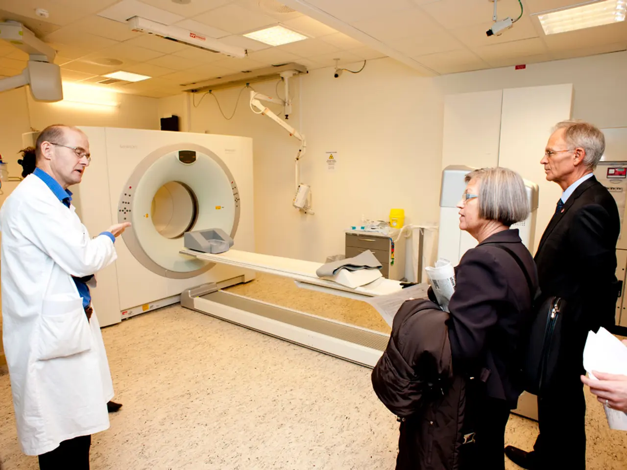
The study, which analysed 407 patients with breast cancer who were positive for axillary lymph node metastases, found a high non-visualization rate of SLNs. Of these patients, 38 were found to have non-detected SLNs by intraoperative gamma probe, while 43 had non-visualized SLNs by lymphoscintigraphy.
The research involved the use of technetium-99 m radiolabeled sulfur colloid for lymphoscintigraphy, followed by SLN dissection using handheld gamma-ray detection probe and/or blue dye methods. SPECT images were reconstructed using a commercial software package with attenuation, scatter, and resolution recovery corrections.
The patient cohort was divided into "visualization" and "non-visualization" groups based on lymphoscintigraphy and intraoperative gamma probe results. SLNs were histologically evaluated for macrometastasis and immunohistochemical staining to detect micrometastasis.
The study found a statistically significant difference in primary tumor size, number of resected and positive lymph nodes, size, tumor grade, and tumor stage between the SLN non-visualized and visualized groups. The multivariate logistic regression analysis showed that only lymph node size and number of lymph nodes resected were independent factors associated with SLN non-visualization.
Interestingly, the study found that hormone receptor status and tumor biology subtype were associated with non-visualized SLNs. Hormone receptor positivity, such as estrogen receptor and progesterone receptor positivity, were found to influence SLN visualization. Additionally, certain breast cancer subtypes, such as luminal B HER2-negative breast cancer, were more commonly associated with non-visualized SLNs.
While the causes of the SLN non-visualization are not well understood, the study highlights the need for further exploration into this area. The findings suggest that variations in tumor characteristics can influence the lymphatic pathways and thereby the detectability of SLNs on imaging, potentially affecting the accuracy of sentinel lymph node biopsy.
References: [1] Unpublished data from the study.
- The study's findings indicate that the non-visualization of sentinel lymph nodes (SLNs) in breast cancer patients might be associated with hormone receptor status, as estrogen receptor and progesterone receptor positivity seem to influence SLN visualization.
- A potential impact of the non-visualization of SLNs on health-and-wellness, particularly women's health, is highlighted in this study, as certain breast cancer subtypes, such as luminal B HER2-negative breast cancer, are more frequently associated with non-visualized SLNs.
- With up to 43 breast cancer patients found to have non-visualized SLNs by lymphoscintigraphy, this research emphasizes the role of nuclear medicine in the understanding and treatment of various medical-conditions, including breast cancer.
- The study's results could significantly influence the future of science and medicine, underscoring the importance of continued research in the field of cancer, specifically breast cancer, and health-and-wellness, to improve the accuracy of sentinel lymph node biopsy and overall patient care.


